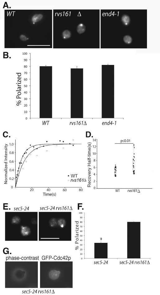
FIGURE 5:
Cdc42p polarization in endocytosis mutants. (A) The wild-type, rvs161Δ, and end4-1 cells were grown to log phase, incubated at 37°C for 90 min, fixed, and immunostained for Cdc42p. Cdc42p is polarized in the endocytosis mutants. Scale bar = 10 μm. (B) Quantification of Cdc42p polarization in wild-type, rvs161Δ, and end4-1 cells. Fifty cells were counted for each group (n = 3). Error bars represent standard error. (C) FRAP curves of GFP-Cdc42p in wild-type and rvs161Δ cells. The normalized fluorescence intensity is plotted over time using SigmaPlot. (D) Recovery half-times for GFP-Cdc42p in wild-type and rvs161Δ cells. Bottom and top of the box are the lower and upper quartiles, respectively. The band near the middle of the box is the median. Whiskers represent standard errors. n = 25, p ≤ 0.01. (E) The sec5-24 and sec5-24 rvs161Δ cells were grown to early log phase, shifted to 35°C for 90 min, fixed, and immunostained for Cdc42p. Although mostly depolarized in the sec5-24 mutant, Cdc42p is well polarized in the sec5-24 rvs161Δ double mutant cells. (F) Quantification of Cdc42p polarization in the sec5-24 and sec5-24 rvs161Δ double mutant cells. Fifty cells were counted for each group (n = 3). Error bars represent standard error. *, p ≤ 0.01. (G) The sec5-24 rvs161Δ cells were transformed with GFP-Cdc42, grown to early log phase, and shifted to 35°C for 90 min. GFP-Cdc42p was then observed using fluorescence microscopy. GFP-Cdc42p is enriched at daughter cell plasma membrane.

