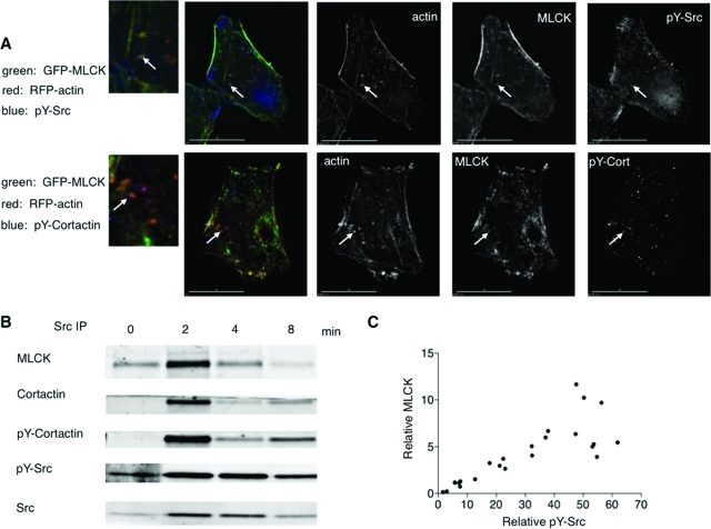
FIGURE 3:
Activated pY-Src and pY-cortactin are found associated with actin/MLCK patches, and immunoprecipitation analysis is consistent with a Src/cortactin/MLCK complex formed on cell swelling. (A) Immunostaining of cells fixed 1 min after hypotonic exposure shows that MLCK colocalizes with pY-Src and pY-cortactin in patches at the base. One patch in each cell is marked with an arrow below it. A magnification of the area of interest is marked with the arrow to the left. (B) MLCK association with activated Src is time dependent after cell swelling. Whole cell lysates (1 mg protein) were prepared from cells that underwent hypotonic exposure for the times indicated (time = 0, initial isotonic conditions), and Src immunoprecipitation was performed. A representative immunoblot of the immunoprecipitates is shown. (C) MLCK and pY-Src signals for each time point (including isotonic controls) were divided by the corresponding fold increase in Src (relative to signal at time = 0), in analogy to methods previously described (Barfod et al., 2005). Correlation analysis of six independent experiments shows that MLCK is significantly associated (Spearman r = 0.8678, p < 0.0001) with pY-Src in Src immunoprecipitates from swollen cells.

