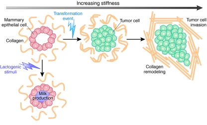
Fig. 2.
Modeling mammary epithelial cells in vitro. Schematic to show modeling of mammary epithelial cells (MECs) in 3D assays in vitro. MECs form organized and polarized acini structures when grown in reconstituted basement membrane in vitro. Milk production can be induced through stimulation with lactogenic hormones. A transformation event results in cell invasion into the lumen. Increased invasive ability correlates with the development of disorganized and branching structures. Increasing matrix stiffness can also induce these events. Adapted, with permission, from Kass et al. (Kass et al., 2007) and Butcher et al. (Butcher et al., 2009).

