Fig. 3.
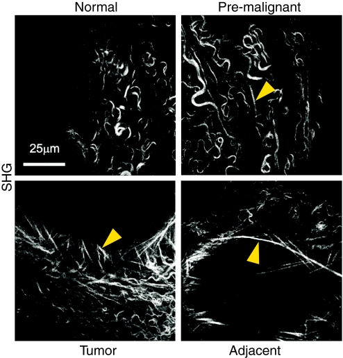
Second harmonic generation (SHG) imaging of collagen fibril linearization during mammary gland tumorigenesis. Images are representative of whole, unfixed mammary glands of MMTV-neu mice [which carry an activated neu oncogene driven by a mouse mammary tumor virus (MMTV) promoter] and show that collagen fibril linearity increases with malignant progression, correlating with increased tissue stiffness. Arrowheads indicate linearized collagen fibrils. Image adapted, with permission, from Levental et al. (Levental et al., 2009).

