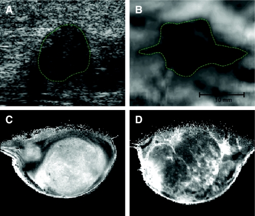
Fig. 4.
Imaging the biomechanical properties of the matrix. Breast tumors are typically identified by changes in tissue mechanics, which can be detected physically, through palpitation or via imaging modalities that exploit tumor-associated changes. (A,B) Images of human breast tumor identified by ultrasound echogram (A) and mammary elastography imaging (B). The dashed lines roughly outline the imaged lesion boundary. The elastogram seems to shows a larger apparent lesion width but a similar height, relative to the echogram. This seems to be due to lateral protrusions, which are consistent with a desmoplastic response associated with local invasion that takes advantage of existing ductal and vascular anatomy. (C,D) MRI images of a 1-methyl-1-nitrosourea (MNU)-induced mammary carcinoma in rat. Here, not only do the changes in tumor ECM provide enhanced tissue contrast, but the intrinsic susceptibility of MRI exploits the paramagnetic properties of deoxyhemoglobin in erythrocytes. Deoxyhemoglobin therefore acts as an intrinsic, blood-oxygenation-level-dependent contrast agent, further highlighting the highly vascular nature of tumors. Ultrasound and elastography images were supplied courtesy of Jeff Bamber (The Institute of Cancer Research, UK). MRI images were supplied courtesy of Simon P. Robinson (The Institute of Cancer Research, UK).

