Fig. 2.
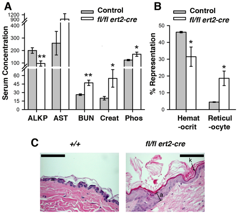
Altered levels of serum biomarkers, anemia and skin abnormalities in induced SHP-2-deficient mice. (A) Shown are the mean levels of the indicated biomarkers in serum ± 1 s.e.m. of tamoxifen-injected (at 6–8 weeks) moribund ptpn11fl/fl ert2-cre mice and littermate control mice (n=6 each genotype). Age range of mice at the time of analysis was 9–12 weeks. ALKP and AST concentrations are shown in U/l; BUN, mg/dl; phosphorous, μg/ml; creatinine, μg/cl. Statistical significance was determined using a two sample Student’s t-test. *P<0.05; **P<0.005. (B) Depicted are mean hematocrit (percent blood volume) and reticulocyte counts (percent blood cells) ± 1 s.e.m. performed on heparinized whole blood from mice in A. Statistical significance was determined using a two sample Student’s t-test. (C) Representative H&E-stained skin section (n=6) of a tamoxifen-injected (at 6 weeks of age) moribund ptpn11fl/fl ert2-cre mouse and a littermate ptpn11+/+ control showing hyperkeratosis (k) and acanthosis (a) in the former. Analysis was performed 4 weeks after tamoxifen injection. Scale bars: 200 μm.

