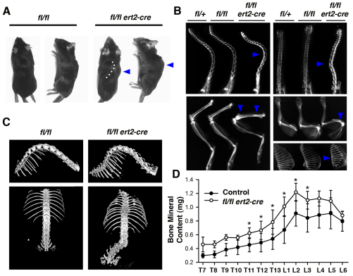
Fig. 4.
Spinal curvature and increased bone mineral content in induced SHP-2-deficient mice. (A) Gross morphology of moribund ptpn11fl/fl ert2-cre mice and ptpn11fl/fl littermate control mice. Note lateral spinal curvature (dotted line) and hump (arrowhead) in the ptpn11fl/fl ert2-cre mouse. (B) X-rays of tamoxifen-injected moribund ptpn11fl/fl ert2-cre mice showing kyphosis (top left) and scoliosis (top right), compared with the indicated littermate controls. Note also increased radiodensity of vertebral bodies of spines, metaphyses of femur (bottom left) and humerus (middle right), and entire rib bones (bottom right) of the ptpn11fl/fl ert2-cre mice (all indicated with arrowheads). In A and B, mice were treated with tamoxifen 5 weeks previously, at 6–8 weeks of age. (C) μCT images of the isosurfaces of spines of ptpn11fl/fl ert2-cre mice and ptpn11fl/fl littermate control mice injected with tamoxifen 5 weeks previously, at 7 weeks of age. Images show scoliosis with rotated vertebral bodies. (D) Mean bone mineral content ± 1 s.e.m. of individual thoracic (T) and lumbar (L) vertebrae from ptpn11fl/fl ert2-cre mice and littermate controls determined by μCT scanning (n=4 for each mouse strain). All mice were treated with tamoxifen 5 weeks previously, at 7 weeks of age. Statistical significance was determined by paired Student’s t-test. *P<0.05.

