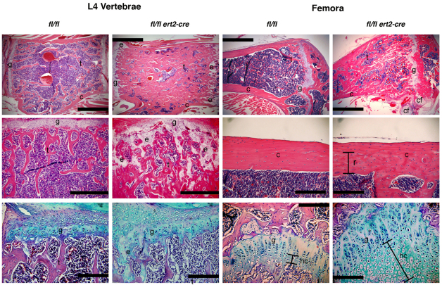
Fig. 5.
Increased and disorganized bone and cartilage in induced SHP-2-deficient mice. Shown are representative images of L4 vertebrae and femora of moribund tamoxifen-injected ptpn11fl/fl ert2-cre mice and ptpn11fl/fl littermate controls. Mice were injected with tamoxifen at 7 weeks of age and analysis was performed at 12 weeks of age. Top and middle panels are stained with H&E, and bottom panels are stained with Alcian blue to highlight cartilage. Scale bars: 1 mm in top panels, 500 μm in L4 vertebrae middle panels, and 200 μm in femora middle panels and all bottom panels. The amount of trabecular bone (t) is dramatically increased in ptpn11fl/fl ert2-cre vertebrae and femora. A region of remodeled (r) cortical bone (c) in femora of ptpn11fl/fl ert2-cre mice shows disorganized bone in this region compared with the same region in the control. Remains of ectopic cartilaginous elements (e) were identified in the trabecular bone region and ectopic cartilage formation (cf) was identified next to growth plates (g) in ptpn11fl/fl ert2-cre mice. The columnar formation of growth plates was disorganized in ptpn11fl/fl ert2-cre mice and the area of hypertrophic chondrocytes (hc) was elongated.

