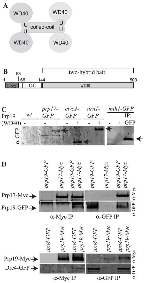
Figure 1. Identification of Prp19 interactors.
A) Model of Prp19 architecture. Domains are not drawn to scale. U = U-box. B) Schematic of Prp19 domains drawn to scale. The region used for the two-hybrid screen is indicated. C-C = coiled-coil. C) Ni2+-NTA beads alone (−) or with (+) bound Prp19(144–503) were incubated with S. cerevisiae protein lysates expressing the indicated GFP-tagged proteins. Bound proteins and anti-GFP immunoprecipitated protein were detected by immunoblotting with anti-GFP antibodies and are indicated with arrows. D) Anti-Myc (left panels) or anti-GFP immunoprecipitates (right panels) from the indicated S. pombe strains were blotted with antibodies to the Myc epitope (top panels) or GFP (bottom panels). Asterisks indicate a band recognized by the anti-GFP antibodies non-specifically in anti-Myc antibody immunoprecipitations.

