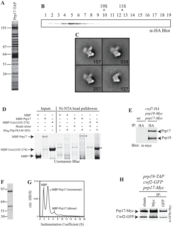
Figure 2. Characterization of Prp19-Prp17 interaction.
A) A fraction of the SpPrp17-TAP eluate was analyzed by silver staining. Positions of markers are indicated. B) SpPrp17-HA3-TAP eluate was resolved on a 10 to 30% sucrose gradient, and fractions were collected from the bottom (fraction1). These were resolved by SDS-PAGE and immunoblotted with anti-HA to detect the migration of Prp17. Migration of of catalase (11.3S) and thyroglobulin (19S) collected from parallel gradients is indicated with asterisks. C) Four representative class averages of SpPrp17-TAP particles in negative stain. The number of particles in each projection average is shown in the lower right corner of each average. Side length of individual panels is 537.6 Å. D) Purified and soluble MBP, MBP-ScUrn(165–274), or MBP-ScPrp17 (Inputs) were incubated with Ni-NTA beads alone or Ni-NTA beads coated with His6-ScPrp19(144–503). Proteins bound to the beads after washing were detected by Coomassie blue staining. Asterisks indicate MBP-ScPrp17 and MBP-ScUrn1 fragments pulled down by the ScPrp19 WD40 domain. The Ni-NTA beads alone did not pull down MBP or MBP fusion proteins, but did pull down some non-specifically binding bacterial proteins. E) An anti-HA immunoprecipitate from S. pombe cwf7-HA prp19-Myc13 prp17-Myc13 cells was blotted for the presence of Myc-tagged proteins. Bands were quantified on an Odyssey instrument. F) Coomassie stained gel of purified MBP-ScPrp17 produced in E. coli. Note the degradation bands. G) Continuous size distribution analysis of sedimentation velocity data of MBP-ScPrp17. AU experiments were conducted at 22°C at a speed of 30,000 rpm and concentration profiles measured at 280 nm. H) SpPrp19-TAP complex was isolated from a S. pombe prp19-TAP cwf2-GFP prp17-Myc13 strain and a portion of the eluate was probed for the presence of SpPrp17 and SpCwf2. The remainder of the eluate was divided in half. One half was immunoprecipitated with anti-Myc and the other with anti-GFP and then each immunoprecipitate was immunoblotted with anti-GFP or anti-Myc antibodies.

