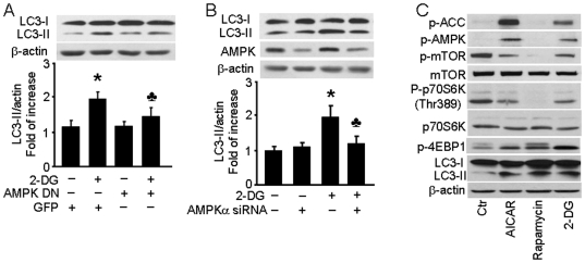
Figure 3. 2-DG-induced autophagy is AMPK-dependent.
A: BAEC were transduced with adenovirus vectors encoding dominant negative AMPK (AMPK-DN) for 48 hrs and then treated with 5 mM 2-DG for 24 hrs (n = 3; two-way ANOVA, * p<0.05, GFP vs. 2-DG, p<0.05, 2-DG vs. 2-DG + AMPK-DN). B: HUVEC were transfected with AMPK-targeted siRNA or control siRNA for 24 hrs then treated with 5 mM 2-DG for 24 hrs. Cell lysates were analyzed by Western blot using antibody against LC3 (n = 3; two-way ANOVA, * p<0.05, vehicle vs. 2-DG, p<0.05, Control siRNA vs. AMPK siRNA). C: BAEC were treated with 1 mM AICAR, 10 nM rapamycin, or 5 mM 2-DG for 24 hrs and then analyzed by Western blot to determine the level of phospho-AMPK-Thr172 (p-AMPK), phos-ACC-Ser79 (p-ACC), phos-mTOR-Ser2448 (p-mTOR), mTOR, phos-p70S6K-Thr389 (p-p70S6K), p70S6K, phos-4EBP1-Thr37/46 (p-4EBP1), and LC3.

