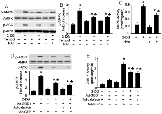
Figure 4. Inhibitory effects of antioxidants on 2-DG-induced AMPK activation in BAEC.
A, B: Confluent BAEC monolayers were pretreated with 10 µM 4-hydroxy-Tempol (Tempol) or 2 mM N-Acetyl-Cysteine (NAc) for 30 min and then treated with 5 mM 2-DG for 10 min. Cell lysates were subjected to Western blot analysis using antibodies against p-AMPK, p-ACC, AMPKα1/2, and β-actin (n = 3; two-way ANOVA,* p<0.05, 2-DG vs. control, Tempol + 2-DG vs. Tempol, NAc + 2-DG vs. Nac, p<0.05 2-DG vs 2-DG + Tem or NAc). C: BAEC were pretreated with NAc and then stimulated with 2-DG. AMPK activity was determined by SAMS phosphorylation using a [32P]ATP assay (n = 3; two-way ANOVA, * p<0.05, vehicle vs 2-DG, Nac vs 2-DG + NAc, p<0.05 2-DG vs. 2-DG + NAc). D, E: Confluent BAEC monolayers were transduced with adenovirus vectors encoding SOD1 or catalase for 48 hrs and then treated with 5 mM 2-DG for 10 min. Phosphorylation of AMPK and AMPK activity were detected, as described in the Matierals and Methods (n = 3; two-way ANOVA, * p<0.05, GFP vs. 2-DG, SOD1 vs 2-DG + SOD1, catalase vs 2-DG + catalase, p<0.05, 2-DG vs. 2-DG + SOD1 or catalase overexpression).

