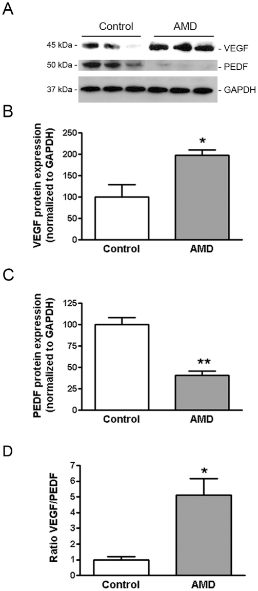
Figure 4. VEGF expression is increased and PEDF expression decreased in RPE from AMD patients.
VEGF and PEDF protein expression was evaluated by Western blot in RPE lysates from 3 smoker donors with AMD and 3 non smoker controls with no known history of eye disease. GAPDH served as loading control. (A) Representative Western blots of the indicated proteins. The numbers to the left are molecular weights in kilodaltons (KDa). (B, C) Average densitometry results. (D) VEGF-to-PEDF protein ratio. Data are expressed as percentage of control and are means ± SE. * is p<0.05 and **p<0.01 versus control.

