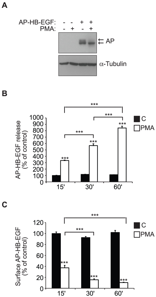
Figure 1. PMA-induced ectodomain shedding of HB-EGF from the cell surface.
A) Western blot analysis of total cell extracts from HT1080 cells stably expressing alkaline phosphatase tagged HB-EGF (AP-HB-EGF) or mock transfected controls after 30 min treatment with 400 nM PMA or DMSO control. α-Tubulin is used as an internal loading control. B) AP-HB-EGF release measured as alkaline phosphatase activity (absorbance at 405 nm) in conditioned media, and C) surface AP-HB-EGF measured as cell surface fluorescence intensity per cell (FLU/cell) in AP-HB-EGF expressing HT1080 cells after 15, 30, or 60 min treatment with 400 nM PMA (white bars) or DMSO control (black bars). The graphs show average values ± standard error of the mean of triplicate experiments. *p<0.05; **p<0.01, ***p<0.001 after one-way ANOVA with Bonferroni's post tests for multiple comparisons. Unless otherwise indicated, the comparison is relative to the respective control.

