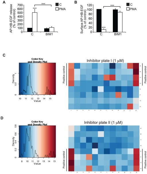
Figure 3. PMA-induced HB-EGF shedding is PKC-dependent.
A) AP-HB-EGF release measured as alkaline phosphatase activity (OD 405 nm) in conditioned media and B) surface AP-HB-EGF measured as cell surface fluorescence intensity per cell (FLU/cell) in DMSO control-treated (C; black bars) and 30 min PMA-treated (PMA; white bars) AP-HB-EGF expressing HT1080 cells treated with the broad PKC inhibitor BIM1 (2 µM) or vehicle control. C, D) Representative heat maps of cell surface fluorescence intensity per cell (FLU/cell) in PMA-treated (400 nM, 30 min) AP-HB-EGF expressing HT1080 cells treated with 1 µM of the InhibitorSelect™ 96-Well Protein Kinase Inhibitor Library I and II, respectively. Color key and density plots are shown to the right of the heat maps. All graphs show average values ± standard error of the mean of at least three independent experiments each done in triplicate. *p<0.05; **p<0.01, ***p<0.001 after one-way analysis of variance with Bonferroni's post tests for multiple comparisons. Unless otherwise indicated, the comparison is relative to the respective control.

