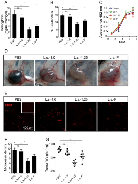
Figure 5. Effect of polysaccharide preparations on tumor growth and angiogenesis in vivo.
C57BL/6 (B6) mice were injected with 500 µl of Matrigel containing 1×10⁵ B16-F10 cells in PBS or 100 µg of a non-fractionated polysaccharide mixture L.s.-P or its fractions L.s.-1.0 and L.s.-1.25. After 6–7 days, tumors were removed and hemoglobin content was evaluated by using the Drabkin colorimetric method. Results are expressed as the amount of hemoglobin (mg)/Matrigel weight (mg) (A) (**P<0.01). (B) Flow cytometry analysis of the frequency of CD34+ endothelial cells on Matrigel plugs embedded with B16 melanoma cells. (**P<0.01) (C) In vitro cell growth of B16 melanoma cells exposed to 100 µg/ml of L.s.-P or its fractions L.s.-1.0 and L.s.-1.25. Data are the mean ± SEM of three independent experiments. (D) B6 mice were injected with 500 µl Matrigel containing 1×10⁵ B16-F10 cells. L.s.-P or its fractions L.s.-1.0 and L.s.-1.25 were injected i.p. at doses of 50 mg/kg every 3 days and compared to control (PBS). Tumors were removed on day 21 post-implantation, photographed (D) and analyzed for CD31+ associated blood vessels (E), microvessel density (F) and weight (G). (*P<0.05; **P<0.01).

