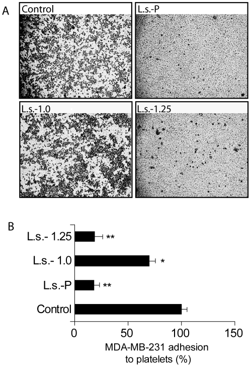
Figure 6. Selective effects of L.s.-P and its fractions L.s.-1.0 and L.s.-1.25 on breast cancer cell adhesion to human platelets.
(A) MDA-MB-231 breast cancer cells were pre-incubated with selected polysaccharide preparations prior to exposure to human platelet-coated plates. The images are representative of three independent experiments. (B) Quantitative analysis of cell adhesion was performed by counting the number of tumor cells adhered to at least three different fields. Results are expressed as mean percentage ± SEM of the treated samples versus control. *P<0.05; **P<0.01.

