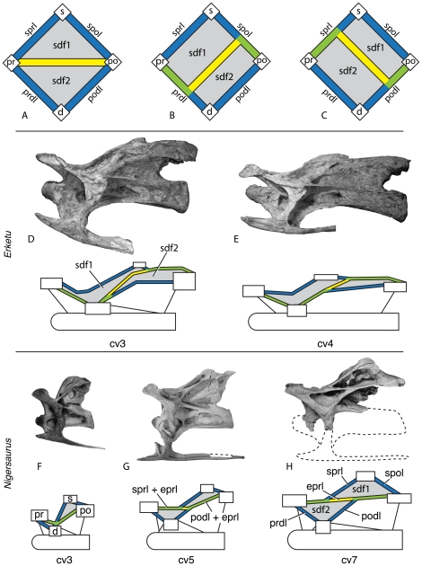
Figure 6. Variable development of the epipophyseal-prezygapophyseal lamina (eprl) and the divided spinodiapophyseal fossa (sdf) in cervical vertebrae.
The eprl in its most basic form (A) connects the prezygapophysis directly with the epipophysis of the postzygapophysis, dividing the sdf into upper (sdf1) and lower (sdf2) subfossae. More commonly, the eprl is conjoined for at least a portion of its length with two or more laminae (B, C), although sdf1 and sdf2 are still readily identifiable. Blue (sprl, spol, prdl, podl) and yellow (eprl) bars represent single laminae; green bars represent conjoined laminae. Examples of conjoined eprl and the indentification of the fossae they bound are given using the holotypic cervical vertebrae of Erketu ellisoni (IGM 100/1803; D, E) and Nigersaurus taqueti (MNN-GAD 512; F–H) in left lateral view, with diagrammatic representation of laminae and landmarks bounding the sdf. Development of the eprl dividing the sdf is dependent on relative position of landmarks, particularly the separation of the summit of the neural spine (s) and the postzygapophysis (po), as well as the relative positions of the prezygapophysis (pr) and diapophysis (d). Even in taxa with a strongly developed eprl, such as Nigersaurus, the lamina is separate from either the sprl or the podl for only a short distance. Taxa with extremely elongate cervical vertebrae, such as Erketu, may have a slightly different arrangement of connectivity between the eprl, spino-zygapophyseal laminae, and zygapophyseal-diapophyseal laminae, although the presence of the eprl can still be traced. Seventh cervical vertebra of Nigersaurus reversed from right lateral. Not to scale.

