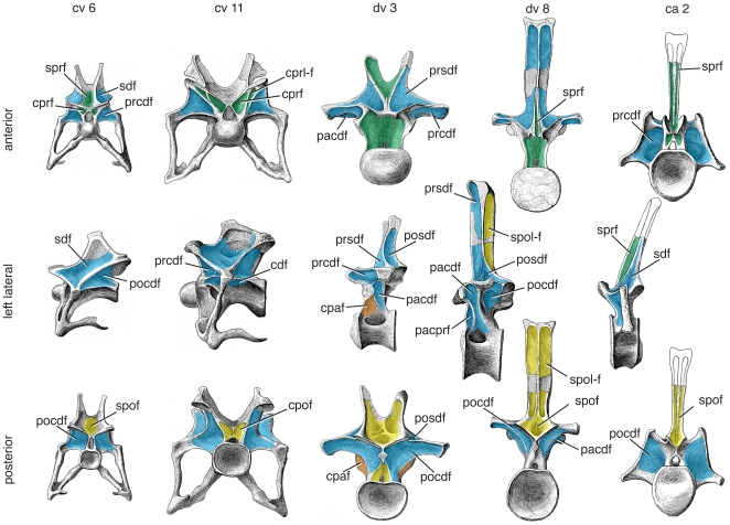
Figure 12. Representative vertebrae of Apatosaurus louisae.
Anterior (top), left lateral (middle), and posterior (bottom) views of anterior cervical (cv 6), posterior cervical (cv 11), anterior dorsal (dv 3), posterior dorsal (dv 8), and anterior caudal (ca 2) vertebrae representing a single individual (CM 3018) and are to scale. Important changes along the column include the loss of the sprf and spof in bifid-spined posterior cervical and anterior dorsal vertebrae, appearance of the prcprf in posterior cervical vertebrae, and the division of the cdf into the cdf and cpaf in mid- and posterior dorsal vertebrae. Images modified from [52]:pls. 24–26). Abbreviations and color scheme as in Figure 7.

