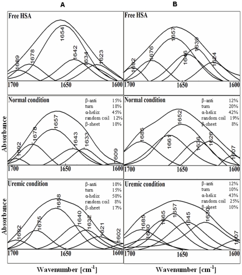
Figure 4. Determination of protein secondary structure complexed with creatinine.
Curve fitted amide I region (1700–1600 cm−1) with secondary structure determination of the free HSA and its toxin adducts in aqueous solution with varying ACR molar ratios and 500 µM protein concentrations at pH 7.0 (A) and pH 9.0 (B).

