
Fig. 6.
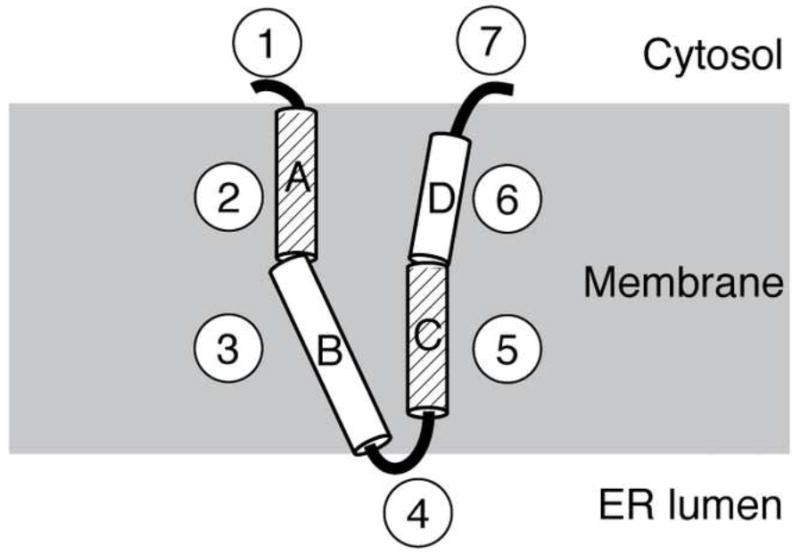
Schematic structural representation of p7 in a phospholipid bilayer. Each of the seven structural segments is shown, with the four helical segments labeled A through D. The dynamic helices, A and C, are differentiated by their diagonal line pattern. The loop containing the two basic residues is shown facing the ER lumen while the N- and C-termini are shown exposed to the cytosol. The helical segments labeled B and D are shown tilted at 25 and 10°, respectively.