
Figure 1. PSEN-1 and YAP Immunohistochemistry in E16, E17, and E18 fetal mouse lungs.
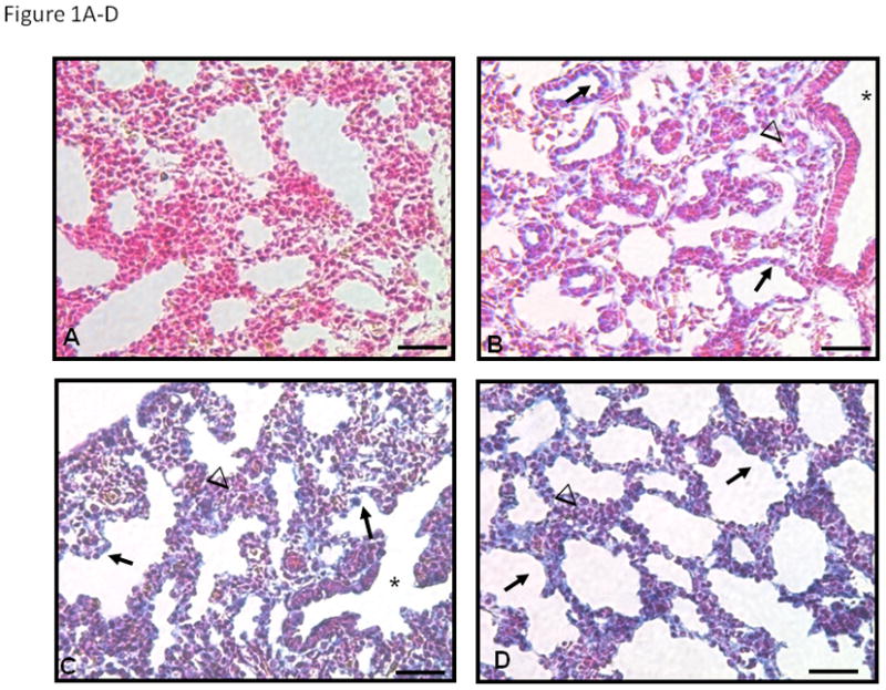
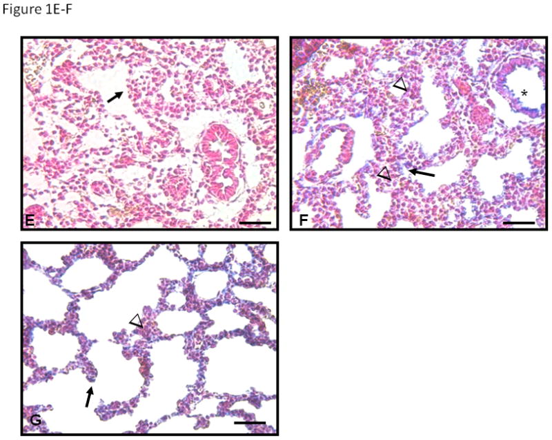
In the absence of primary antibody (A) no blue staining is seen. At E16 (B) PSEN-1 protein is seen in the cytoplasm of the cuboidal epithelium (arrows in B) and in surrounding mesenchyme (arrowheads). However columnar epithelia of bronchiolar airways (asterisk) are negative. Expression of PSEN-1 at E17 (C) and E18 (D) increases in cuboidal epithelium (arrows) and epithelium of distal bronchioles (asterisk in C) at the entrance to alveolar ducts. Mesenchymal cells adjacent to cuboidal epithelium are also strongly positive but clusters of mesenchymal cells between airways (arrowheads in C and D) are less positive, specifically in E18 (D). YAP cellular expression is minimal at E16 (E), but at E17 (F) and E18 (G) expression is present in cuboidal epithelium (arrows) particularly at tips of alveolar ducts. Low cuboidal epithelium (asterisk) is also positive at E17 and E18. Minimal YAP staining is present in mesenchyme (arrowheads in F and G). Bars are 50 microns.