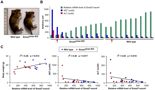
Figure 2. Deletion of Smad7 causes spontaneous liver failure.
(A) Representative images of 12-week-old wild type and Smad7liver-KO mice. (B) Relative mRNA level of Smad7 exon4 (green) as comparison with the serum AST (blue) and ALT (red) values. The arrows indicate the mice that were used in histological and immunohistochemical analyses performed in Figure 3. (C) Correlation analyses of body weight, AST and ALT levels with the mRNA level of Smad7 exon4. The values of body weight, AST and ALT values were plotted against the relative mRNA level of Smad7 exon4 for each mouse (red for Smad7liver-KO mice and blue for wild type mice) and subjected to linear regression analysis. n = 11 for Smad7liver-KO and n = 10 for wild type mice.

