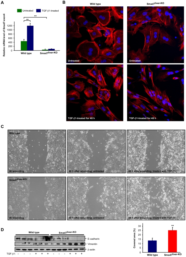
Figure 4. Deletion of Smad7 enhances TGF-β-induced EMT.
(A) Confirmation of Smad7 deletion in primary hepatocytes isolated from Smad7liver-KO mouse. Real time RT-PCR was performed with total RNA isolated from wild type or Smad7liver-KO mice with primers to detect the mRNA region corresponding to exon4 of Smad7 gene. The data are shown as mean ± SD and ** indicates p<0.01 as comparison between the groups as indicated by Student's t-test. (B) TGF-β-induced EMT-like morphology changes. Immunofluorescence labeling were performed with wild type or Smad7liver-KO hepatocytes treated with or without TGF-β1 (5 ng/ml) for 48 h. F-actin was stained with fluorescein isothiocyanate-labeled phalloidin (Red) and the nuclei were labeled by Hoechst 33342 (Blue). (C) Analysis of cell motility by a wound-healing assay. Cultured primary hepatocytes were analyzed by phase contrast microscopy. The cells were treated with or without TGF-β1 (5 ng/ml) for 48 h. Quantitation of the cell motility is shown in lower right panel as mean ± SD and ** indicates p<0.01 by Student's t-test. (D) Analysis of EMT markers E-cadherin and vimentin. Primary hepatocytes were treated with or without TGF-β1 (5 ng/ml) for 48 h and the cell lysate was used in immunoblotting with the antibodies as indicated.

