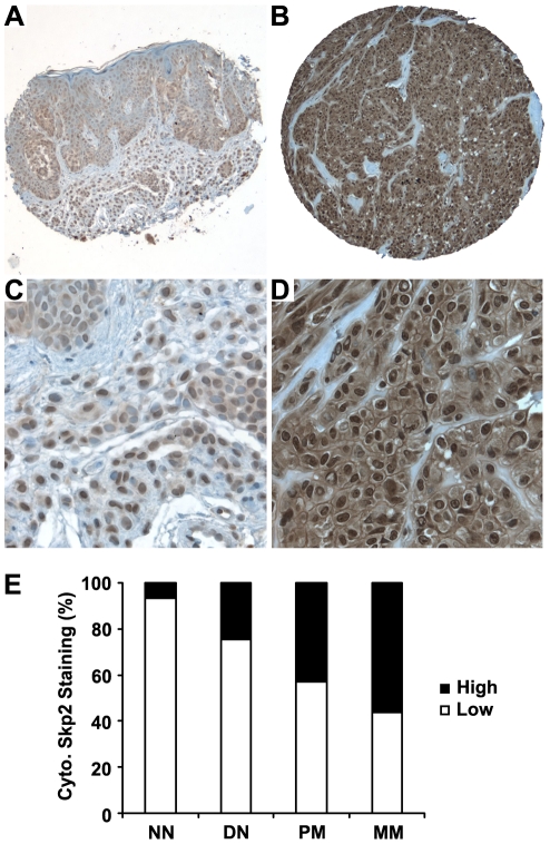
Figure 2. Cytoplasmic Skp2 expression is increased in human melanomas.
Representative images of cytoplasmic Skp2 immunohistochemical staining in human melanocytic lesions. (A and C) Low cytoplasmic Skp2 staining, (B and D) High cytoplasmic Skp2 staining. (E) Cytoplasmic Skp2 expression is increased from normal nevi to dysplastic nevi and melanoma. Significant differences for Skp2 staining are observed between normal and dysplastic nevi (P = 0.039, χ2 test), dysplastic nevi and primary melanoma (P = 0.008, χ2 test), and primary and metastatic melanoma (P = 0.008, χ2 test). NN, normal nevi; DN: dysplastic nevi; PM: primary melanoma; MM: metastatic melanoma. Magnification: ×100 for A and B; ×400 for C and D.

