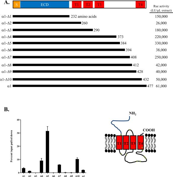
Figure 1. Recombinant AChR-α1 proteins used in LIPS.
(A) The full length and 10 deletion mutants of the α1 subunit of the AChR were cloned into the pREN3S vector to generate N-terminal fusions with Renilla luciferase. SP: signal peptide; ECD: extracellular domain; T: transmembrane segment. Typical recoverable Ruc activity for each construct is shown to the right as LU/μL extract. (B) The D6 monoclonal antibody, directed at the AChR-α1 subunit was used to immunoprecipitate the AChR-α1-Ruc fusion proteins. The amount of protein immunoprecipitated is expressed as a percentage of the total input for each assay. The inset shows a schematic of the AChR-α1 subunit within the plasma membrane; the dots on the figure denote the approximate location of each truncation mutant.

