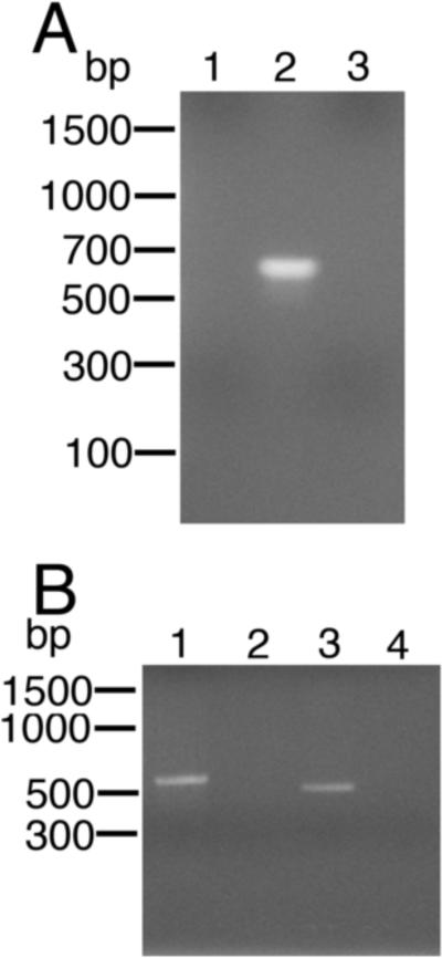
Fig. 7.
PCR and spliced leader (SL) reverse transcriptase (RT)-PCR analysis of progeny microfilariae (mf) recovered from animals co-injected with L3s and calcium phosphate - pBmHSP70/Gluc co-precipitates. A) PCR analysis of DNA extracted from progeny mf utilizing primers derived from the GLuc open reading frame (ORF). PCRs were carried out as described in Materials and methods. Lane 1 = PCR using DNA extracted from mf recovered from an animal injected with L3s alone. Lane 2 = PCR using DNA extracted from mf recovered from an animal co-injected with L3s and calcium phosphate - pBmHSP70/GLuc co-precipitates. Lane 3 = PCR negative control. B) Hemi-nested SL mediated RT-PCR analysis of mRNA extracted from mf utilizing primers derived from the GLuc ORF and the B. malayi SL. Lane 1 = SL mediated RT-PCR positive control (RNA extracted from embryos transfected with pBmHSP70(E1-I1-E2-I2-E3)/luc and amplified using primers derived from the BmHSP70 gene as previously described (Liu et al., 2007)). Lane 2 = RNA extracted from mf recovered from an animal injected with L3s alone. Lane 3 = RNA extracted from mf recovered from an animal co-injected with L3s and calcium phosphate - BmHSP70/GLuc co-precipitates. Lane 4 = RT-PCR negative control.

