
Figure 8. Tregs from TMEV-infected SJL/J and C57BL/6 mice are equally able to suppress proliferation of CD4+ T cells.
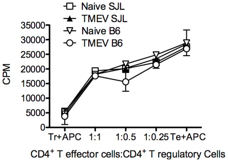
B6-GFP-Foxp3 and SJL/J-GFP-Foxp3 animals were i.p. infected with 1.5×10⁷ pfu/ mouse TMEV. 7 days later splenocytes were isolated and CD4+GFP+ cells were sorted from naïve and infected SJL/J and B6 animals. These cells were cultured with CD4+CD25−Foxp3− cells from naïve (SJLxB6)F1 mice in the presence of 0.2 μg/well anti-CD3 (clone 2C11) using T cell-depleted F1 splenocytes as antigen presenting cells. Cultures were pulsed with 3H-TdR at the time of plating and cultured at 37° C for three days before harvesting. Data is representative of 2–3 individual experiments.