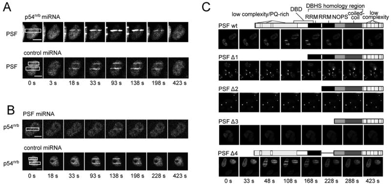
Fig. 3.

Requirement of PSF N-terminal sequences for relocalization of p54nrb to sites of DNA damage. (A, B) HeLa cells were transfected with miRNA-expressing plasmids and transfected again at 24 h with tagged protein expression vectors as indicated. Laser microirradiation and imaging were performed as in Fig. 2C and 2D. Scale bar is 10 μm. (C) Analysis of PSF mutants. Schematic diagrams are as in Fig. 1. Transfection, laser microirradiation and imaging were performed as in Fig. 2. All constructs are PSF-AcGFP fusions except PSF Δ3, which is PSF-dsRed.
