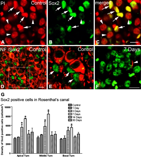
FIG. 9.
Increased Sox2+ cells within Rosenthal’s canal in injured auditory nerves. A–C Nuclear staining pattern of Sox2 antibody (green) in normal auditory nerve. PI nuclear counterstaining (red) reveals nuclear profiles of Sox2+ cells. The nuclei of SGNs are identified by their large spherical nuclei (arrowheads). Note that SGNs do not express or express a very low level of Sox2 and were not considered as Sox2+ cells in this study. D, E Dual immunostaining for Sox2 (green) and a neuronal marker, NF 200 (NF, red) reconfirms that high-intensity expression of Sox2 appears in the nuclei of Schwann- and or satellite-like cells (arrows). E Enlarged image of corresponding boxed area in D. F Sox2+ cells in the auditory nerve 7 days after ouabain treatment. An arrowhead indicates a surviving SGN process. G Mean density of Sox2+ cells within the Rosenthal’s canal in the apical, middle, and basal turns from control, 1-, 3-, 7-, 14-, and 30-day post-treated ears (n = 3–6 per group). Scale bars, 8 μm in C (applies to A–C); 7 μm in D; 7 μm in F (applies to E–F).

