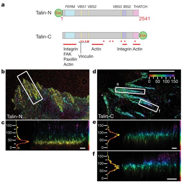
Figure 3. Talin orientation in focal adhesions.
a, Schematic diagram, with important domains and binding sites indicated for Talin PA-FP fusions (FERM, protein 4.1, ezrin, radixin, moesin domain; VBS, vinculin binding sequence; IBS, integrin binding site). Talin-N, N-terminal fusion; Talin-C, C-terminal fusion (Supplementary Table 3). b–f, Top view and side view iPALM images of focal adhesions (white boxes, top-view panels) and corresponding z histograms and fits for talin-N–tdEos (b, c) and talin-C–tdEos (d–f). Colours: vertical (z) coordinate relative to the substrate (z=0nm, red). Scale bars: 5 μm (b, d) and 500 nm (c, e, f).

