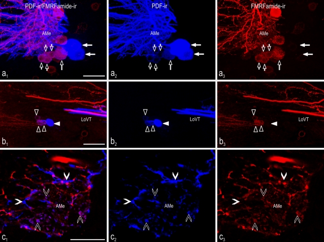
Fig. 2.
Colocalization of PDF and FMRFamide immunoreactivity in neurons and neuropil of the accessory medulla (AMe) of the cockroach. Confocal laser scan images were obtained from vibratome sections of the accessory medulla showing PDF-immunoreactive (blue) and FMRFamide-immunoreactive (red) neurons. Left Overlay images. a, b Maximum projections from stacks of optical sections. c Single optical section. a 1-3 Neurons of the small (small open arrowheads), medium-sized (large open arrowhead), and large anterior PDFMe (large filled arrows). b 1-3 Posterior PDFMe comprising small (open triangles) and large (filled triangles) cells. All small, but no large, posterior PDFMe showed colocalized PDF/FMRFamide immunoreactivity (LoVT lobula valley tract). c 1-3 In the AMe, weakly labeled PDF-ir varicosities (open arrowheads) in the noduli showed colocalized PDF/FMRFamide immunoreactivity, whereas large and intensely labeled PDF-ir varicosities (filled arrowheads) in the internodular neuropil did not. Bars 50 μm (a), 25 μm (b), 100 μm (c)

