Fig. 3.
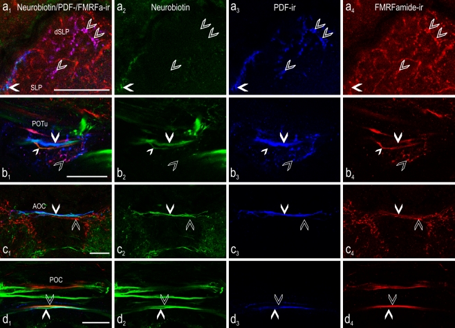
Colocalization of PDF (blue) and FMRFamide (red) immunoreactivity in the central brain of the cockroach combined with neurobiotin (green) backfill from a cut optic lobe stump. Confocal laser scan images (single optical sections) of typical projection areas of the PDFMe in the central brain after backfill of neurobiotin from the cut left optic stalk. Left Overlay images. a 1-4 At the dorsal part of the superior lateral protocerebrum (dSLP) contralateral to the backfilled side, fibers were visible with colocalized PDF and FMRFamide immunoreactivity (open arrowheads), but these were not labeled by neurobiotin. At the distal rim of the superior lateral protocerebrum (SLP) fibers were located showing colocalization of neurobiotin backfill and strong PDF immunoreactivity (filled arrowheads) but they did not exhibit FMRFamide immunoreactivity. b 1-4 In the posterior optic tubercle (POTu), fibers were present with colocalized PDF and FMRFamide immunoreactivity (open arrowheads), but these fibers were never labeled by neurobiotin backfills. In contrast, neurobiotin was found in commissural fibers either immunoreactive to anti-PDF (large filled arrowhead) or to anti-FMRFamide (small filled arrowhead) alone. c 1-4 In the anterior optic commissure (AOC), PDF-ir fibers labeled by neurobiotin backfill were frequently visible (filled arrowheads). These were never additionally colabeled by FMRFamide immunoreactivity, and parallel FMRFamide-ir fibers (open arrowheads) were never labeled by neurobiotin backfill. d 1-4 In the posterior optic commissure (POC), many backfilled fibers crossed the midline of the central brain. Backfilled fibers that showed either colocalized neurobiotin and PDF immunoreactivity (filled arrowheads) or neurobiotin and FMRFamide immunoreactivity (open arrowheads) were often found, but triple labeling was never observed. Bars 50 μm

