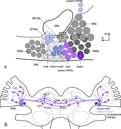
Fig. 6.
a Summary representation of the composition of ipsi- and contralaterally projecting PDF-expressing medulla neurons at the accessory medulla (AMe) of the cockroach. Although the numbers of gray circles do not reflect the actual group sizes, numbers of colored circles represent the mean numbers of the respective PDFMe neurons as determined in this work. All small and medium-sized anterior PDFMe and all small posterior PDFMe appear to coexpress peptide(s) of the FMRFamide-related peptide family. Additionally, all medium-sized PDFMe appear to coexpress an Asn13 orcokinin-like peptide. Asn13-orokinin-like immunoreactivity is also present in at least a subpopulation of the small anterior PDFMe, but not in the small posterior PDFMe. However, all large anterior and large posterior PDFMe do not coexpress one of the tested peptides after PDF. Thus, possibly all medium-sized anterior PDFMe and at least a subpopulation of the small anterior PDFMe express three different neuropeptides simultaneously. Together, six anterior and posterior large (including the largest) PDFMe comprise a homogeneous group of neurons that appear to be randomly distributed to anterior or posterior positions during the cockroach’s development; hence, all large PDFMe are here depicted among the anterior AMe neuron groups. The neurobiotin backfills suggest that the largest and three medium-sized PDFMe project to the contralateral AMe for mutual pacemaker coupling (C). Furthermore, an additional contralaterally projecting VNe neuron appears to exist that is not immunoreactive to one of the peptide antisera tested (DFVNe distal group of frontoventral neurons, MFVNe medial group of frontoventral neurons, MNe medial neurons, VNe ventral neurons, VMNe ventromedial neurons, VPNe ventroposterior neurons, di distal, do dorsal). b Representation of pathways and output regions of contralaterally projecting PDFMe (violet lines pathways of the largest PDFMe, blue lines pathways of the contralaterally projecting medium-sized PDFMe, violet/blue circles respective output regions in the neuropil). The large PDFMe appears to connect the two AMae via the anterior and posterior optic commissure (AOC, POC) and terminates in most output regions of the PDF-ir fiber system. Medium-sized contralateral PDFMe terminate at least in the AMe, the dorsal lateral protocerebrum (dSLP), and the posterior optic tubercles (POTu), as judged by the presence of anti-PDF/FMRFamide colabeled terminal arborizations. The medium-sized contralaterally projecting PDFMe appear to project only via the AOC (La lamina, Me medulla, ILP/SLP inferior/superior lateral protocerebrum, SMP superior median protocerebrum)

