Figure 6.

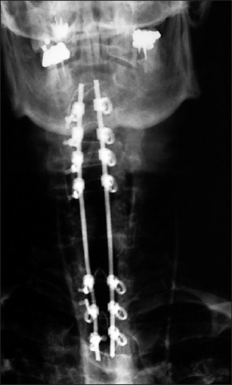
This AP X-ray, following a laminectomy of C5 and C6 demonstrates a rod/eyelet fusion construct involving the C2, C3, C4 and the C7, T1, and T2 levels. The eyelets are located ventrally, while the crimped wires and rods are found dorsally.

This AP X-ray, following a laminectomy of C5 and C6 demonstrates a rod/eyelet fusion construct involving the C2, C3, C4 and the C7, T1, and T2 levels. The eyelets are located ventrally, while the crimped wires and rods are found dorsally.