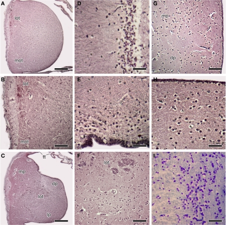
Figure 2.
Photomicrographs of transverse sections through the forebrain of the Comoran coelacanth showing various cell groups. (A,B) The rostral body. (C) The rostral pallium. (D) The striatum. (E) The ventral pallium. (F) The core and cellular band of the lateral pallium. (G) The medial border of the dorsal and medial pallia. (H) The cells of the medial pallium. (I) The cells of the central amygdalar nucleus. Sections in (A–H) are stained by the Bodian silver method; section I is stained with 1% cresyl violet. Scale bars = 100 μm (D,E,H,I), 200 μm (B,F,G), 500 μm (A,C). dp, Dorsal pallium; lot, lateral olfactory tract; lp, lateral pallium; mot, medial olfactory tract, mp, medial pallium; tt telencephalic tela.

