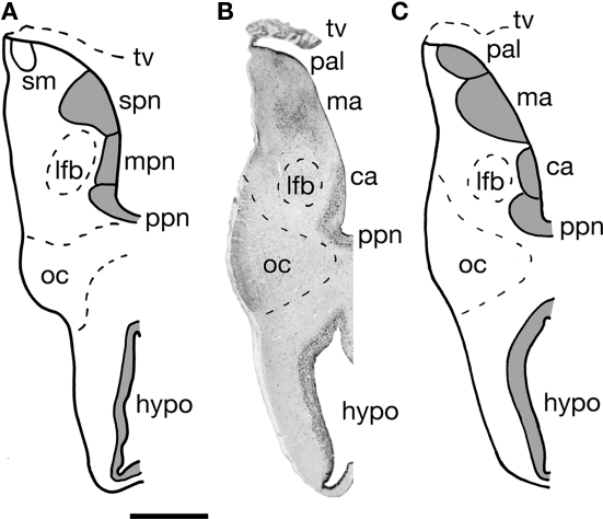
Figure 3.
A transverse section through one half of the telencephalon impar of the Comoran coelacanth. (A) Cell groups as interpreted by Nieuwenhuys (1965). (B) A Nissl-stained section from the present study showing the histology. (C) Cell groups as interpreted in the present study. Bar scale = 1 mm. ca, Central amygdalar nucleus; hypo, hypothalamus; lfb, lateral forebrain bundle; ma, medial amygdalar nucleus; mpn, magnocellular preoptic nucleus; oc, optic chiasm; pal, pallium; ppn, parvocellular preoptic nucleus; stria medullaris; spn, superior preoptic nucleus; tv, transverse velum.

