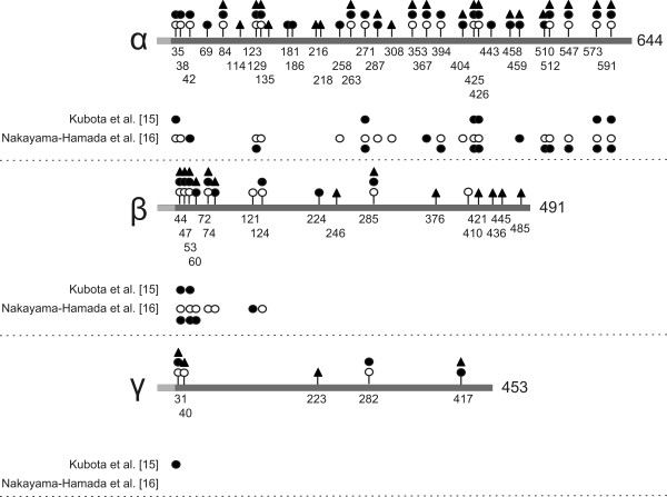
Figure 3.
Citrullination sites in human fibrinogen. Human fibrinogen was citrullinated in vitro by hPAD2, hPAD4 or rmPAD2 and citrullinated residues were determined by proteolytic digestion followed by mass spectrometry. The positions of the citrullination sites are schematically depicted in the three polypeptide chains of fibrinogen: hPAD2, filled circles; hPAD4, open circles; rmPAD2, filled triangles. The grey boxes at the N-termini of the fibrinogen chains represent signal peptides (for all chains) and fibrinopeptides (for the α- and β-chains). Beneath each fibrinogen chain (α, β or γ), previously detected citrullination sites have been depicted; hPAD2, filled circles; hPAD4, open circles [15,16]. Amino acid numbering is relative to the start of the signal peptides.

