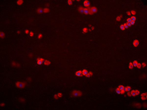
Figure 4.
The presence of chemerin in sections of human articular cartilage. The micrograph (40X) shows positively (red) stained chondrocytes in tissue from one patient undergoing ligament repair. Nuclei were visualized by Dapi dye (blue). Negative controls (isotype IgG) had no red staining (not shown).

