
Figure 6.
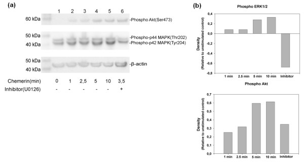
Western blot of phosphorylated p44/42 MAPKs (Thr202/Tyr204) and phosphorylated Akt (Ser 473). (a) Cultured chondrocytes were challenged with 10 nM chemerin21-157 for 1, 2.5, 5 and 10 minutes. Lane 1 represents the control where no chemerin was added and Lane 6 represents the sample extract where the MEK 1/2 kinase inhibitor U0126 was added 1 h prior to a 3.5 minutes chemerin21-157 challenge. (b) The density of each band was normalized to β-actin, the graphs shows the increase in density relative to unstimulated control.