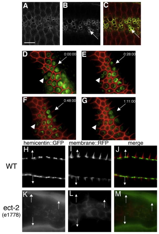
Figure 1. Hemicentin Assembles at the Cleavage Furrow of C. elegans Germ Cells and Prevents Membrane Retraction.
Germ cells in the hermaphrodite distal gonad have incomplete cleavage furrows.
(A–C) Optical section through the central region of C. elegans gonad showing wild-type C. elegans hermaphrodite germ cells coexpressing RFP::phospholipase C δ PH domain (A and C) and hemicentin::GFP (B and C). Note that hemicentin::GFP accumulates at the periphery of incomplete cleavage furrows (arrows).
(D–G) Progressive retraction of germ cell membranes in the absence of hemicentin. Images show a time course of a single him-4(rh319) hermaphrodite mutant gonad expressing RFP:PH and histone 2B:GFP transgenes. Images were collected at 0 (D), 28 (E), 48 (F), and 71 min (G). Note progressive retraction of the germ cell membrane (arrow). Arrowhead indicates one of several multinucleate germ cells present prior to image collection. This effect does not depend on osmotic pressure (Figure S1). Phenotype quantitation is presented in Table S1.
(H–M) The ECT-2 RhoGEF is required for hemicentin assembly in the gonad. Comparison of membrane RFP:PH and hemicentin:GFP distribution in a wild-type and ect-2 mutant background is shown. In a wild-type background, hemicentin:GFP assembles peripheral to incomplete cleavage furrows. In an ect-2(e1778) mutant background, cleavage furrows do not form properly, and hemicentin does not assemble at membrane surfaces but accumulates outside the gonad (arrows). Scale bar represents 5 μm.

