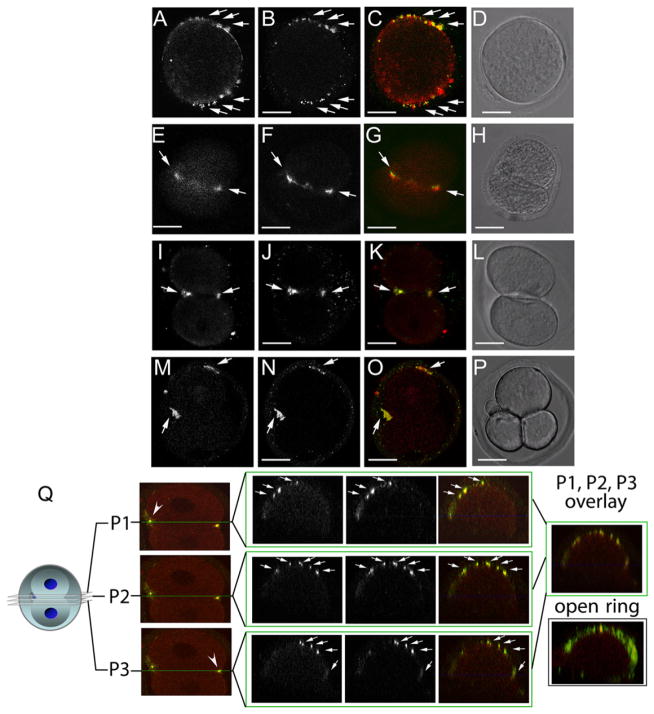
Figure 2. Hemicentin Distribution in Wild-Type Mouse Embryos.
(A–P) Immunofluorescence images of dividing one- (A–D), two- (E–L), and three-cell (M–P) wild-type embryos. Embryos were stained at multiple time points with antibodies to hemicentin (A, E, I, and M) and myosin IIB (B, F, J, and N). Merged images (C, G, K, and O) indicate that hemicentin staining accumulates adjacent to myosin II staining in dividing cells. Also shown are corresponding differential interference contrast (DIC) images (D, H, L, and P). Scale bars represent 20 μm.
(Q) Confocal image of three separate planes across the cleavage furrow of a dividing one-cell embryo stained with hemicentin (red) and myosin IIB (green) antibodies. Right: overlay of merged images from two different embryos showing that both proteins form a punctate open ring around the periphery of the cleavage furrow.

