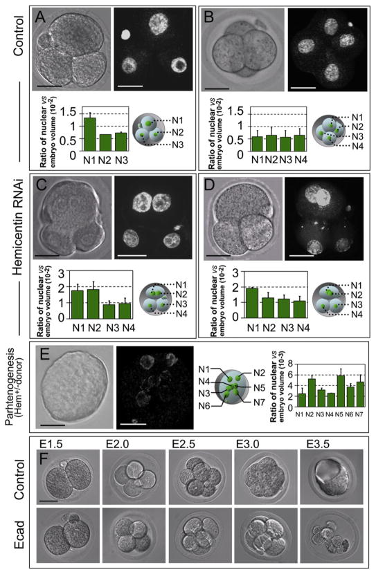
Figure 3. Hemicentin-1 Depletion Results in Embryonic Arrest Prior to Implantation.
(A–D) Wild-type embryos were mock injected (A and B) or injected at the one-cell stage with hemicentin-1 dsRNA (C and D). Control embryos progress through the three- and four-cell stage with single nuclei (A and B). In contrast, hemicentin-1 dsRNA-injected embryos arrest prior to the four-cell stage with multinucleate cells (C and D). DIC and corresponding YOYO-1-stained images are shown. Ratio of nuclear volume to embryonic volume (×10−2) with corresponding schematic diagram is shown below each embryo. Error bars represent standard deviation. Larger nuclear volumes of N1 and N2 in (C) and N1 in (D) suggest that these nuclei are in G2 phase [27].
(E) Parthenogenic activation of eggs obtained from heterozygous hemicentin-1 females results in arrest of embryos with multinucleate cells. DIC and corresponding YOYO-1-stained images of embryos obtained from parthenogenic activation of eggs collected from heterozygous females are shown. Approximately 40% of eggs divide to become mononucleate cells, and ~60% (Table S2) arrest at the one- to three-cell stage, with most cells containing multiple nuclei. At least seven distinct nuclei (N1–N7) are detected within the single arrested blastomere. Ratio of nuclear volume to embryonic volume (×10−3) with corresponding schematic diagram is shown to the right. Error bars represent standard deviation.
(F) As described above, the majority of mock-injected embryos successfully develop into blastocysts (see Table S2). As a positive control, most E-cadherin dsRNA-injected embryos proliferate normally from the two- to eight-cell stage but fail to form a blastocoele and arrest at the morula stage (Table S2), consistent with published information on the phenotype of E-cadherin knockout mice [29].
Scale bars represent 20 μm.

