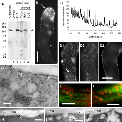
Figure 5.
Characterization of CesA in tobacco pollen tubes. A, Immunoblot with anti-CesA antibody on Arabidopsis extract (20 μg; lane 1), cytosolic proteins (20 μg; lane 2), membrane proteins (20 μg; lane 3), Triton-solubilized cell wall proteins (10 μg; lane 4), NaCl-solubilized cell wall proteins (10 μg; lane 5), and SDS-solubilized cell wall proteins (5 μg; lane 6) from tobacco pollen tubes. The arrowhead indicates the main cross-reacting band around 125 kD in the Arabidopsis, membrane, Triton-, and NaCl-extracted proteins. B, Immunolocalization of CesA in tobacco pollen tubes. The image was captured in the center of the pollen tube. Staining was mainly observed in the apical domain (arrow) and to a lesser extent in the remaining cortical region (arrowhead). In the apex, staining was also diffusely observed in the cytoplasmic domain (asterisk), presumably in association with vesicular material. C, Measure of fluorescence intensity along the cell border of pollen tubes emphasizing the higher signal in the apex and the progressive decline toward a basal level. The presence of numerous peaks indicates the spot-like distribution of CesA proteins. D, A single pollen tube observed at three different focal planes: D1, tangential to the surface (presumably plasma membrane and cell wall); D2, just below the plasma membrane; D3, in the middle. CesA was distributed differently. The arrow indicates invisible tracks along which CesA spots align. Bar = 10 μm. E and F, Double immunolocalization of microtubules (green) and CesA (red). CesA proteins appeared as dots sometimes coaligning with microtubules but also distributed between microtubule bundles. Bars = 5 μm. G, Immunogold labeling of CesA. The protein was detected in association with the plasma membrane (arrows), with vesicular structures below the plasmalemma (arrowheads) and with Golgi bodies (bordered arrow). Bar = 250 nm. H to J, Immunogold labeling of CesA in the cortex of pollen tubes. CesA was mainly found in association with the cortical region of tobacco pollen tubes, presumably with the plasma membrane (H; arrow). Gold particles were frequently found in association with vesicular structures (asterisks) of around 100 to 150 nm that accumulate in proximity of the plasma membrane (I and J). Bars = 250 nm.

