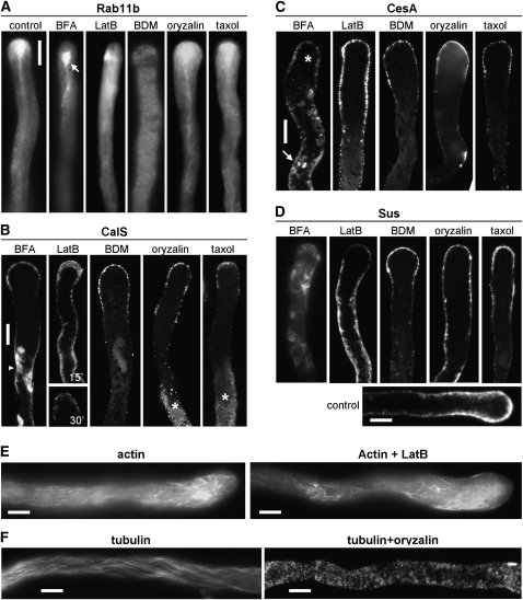
Figure 9.
Distribution of apical vesicles, CalS, CesA , and Sus after treatment with inhibitors. Vesicles were visualized in tobacco plants expressing a GFP-labeled Rab11b, while CalS, CesA, and Sus were visualized in wild-type tobacco plants. Experimental conditions were as follows: 5 μg mL−1 BFA, 2 nm LatB, 30 mm BDM, 1 μm oryzalin, and 10 μm taxol. A, GFP-labeled Rab11b showing the distribution of apical vesicles (the arrow in the second image indicates the BFA-induced membrane compartment). Bar = 10 μm. B, Distribution of CalS after treatment with inhibitors. BFA caused the accumulation of intracellular deposits of CalS (arrowhead), probably corresponding to BFA-induced aggregates. The three-patterned distribution of CalS was maintained but was less apparent. LatB affected significantly the distribution of CalS after 15 min (top panel) and more strongly after 30 min (bottom panel), causing a uniform distribution of CalS in the cortical region and the disappearance of the strong deposits in the apex. BDM treatment allowed the persistence of lower levels of CalS in the apex but removal of CalS in more distal regions of pollen tubes. Oryzalin and taxol did not interfere with the apical distribution of CalS but generated the progressive disappearance of the distal labeled area and the accumulation of intracellular deposits (asterisks). Bar = 10 μm. C, Effects of inhibitors on the distribution of CesA. BFA induced the accumulation of labeling in the cytoplasm and a consistent decrease in the apical region of pollen tubes (asterisk). LatB induced a progressive relocation of the CesA signal toward basal regions, although labeling was still organized as a tip-base gradient. The myosin inhibitor BDM caused a decrease in the accumulation of CesA in the apical/subapical regions and the progressive disappearance of CesA in distal regions. Oryzalin and taxol caused no significant changes in the distribution of CesA, which was detected according to the tip-base gradient. Bar = 10 μm. D, Effects of inhibitors on the distribution of Sus. BFA induced the progressive disappearance of Sus from the tube shanks and its accumulation in the cytoplasm. LatB induced the progressive disappearance of Sus in the apical domain and accumulation in the base region, with signal that gradually occupied the tube cytoplasm. BDM treatment caused the persistence of an evident staining in the apex but a progressive disappearance or absence in the shanks of pollen tubes. Oryzalin and taxol did not have significant effects on the distribution of Sus. The control pollen tube showed the typical cortical distribution of Sus, with more intense signal in the apex. Bar = 10 μm. E, Effects of the AF inhibitor LatB. In control pollen tubes (left panel), AFs are typically organized as bundles in distal regions and as a network in the subapical domain. After treatment with LatB (right panel), bundles of AFs are still present distally but the subapical network disappears. Images are single cortical optical sections. Bars = 10 μm. F, Effects of the MT inhibitor oryzalin on the organization of MTs in the pollen tube. In controls (left panel), MTs show the typical filamentous arrangement, which is lost after treatment with oryzalin (right panel), leaving only diffused fluorescent spots. Images are whole cell projections. Bars = 10 μm.

