Figure 3.
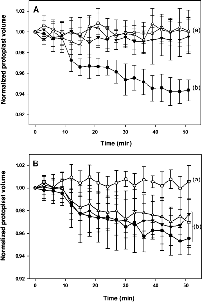
Cell-shrinking assay using motor cell protoplast of S. saman. Protoplasts were prepared from separated half segments of tertiary pulvini of S. saman, i.e. from the extensor or flexor side, respectively. A, LCF initiates shrinkage only in protoplasts derived from extensor cells but not flexor cells. The volume of extensor cell protoplasts (black circles) or flexor cell protoplasts (black triangles) was monitored for 51 min after treatment with (−)-LCF (100 μm) and compared to the untreated control cells (white circles and white triangles, respectively). B, (−)-LCF was applied at different concentrations to extensor cell protoplasts and their shrinking was monitored for 51 min; 100 μm (−)-LCF (black circles), 10 μm (−)-LCF (black triangles), 1 μm (−)-LCF (white triangles), and water control (white circles). The values represent the normalized mean volume (±sd) of six to 12 protoplasts. Different letters indicate significant differences between the terminal measurement (i.e. t = 51 min; Student-Newman-Keuls post-hoc test: P < 0.05, after univariate ANOVA with treatment as fixed factor and time as random factor: A, F3,25 = 26.99; B, F3,21 = 7.47; P < 0.001).

