Figure 5.
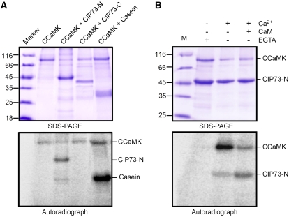
Phosphorylation of CIP73 by CCaMK in vitro. A, Autophosphorylation and phosphorylation assay of CCaMK in the presence of Ca2+ and CaM. CCaMK can phosphorylate CIP73-N (1–413) but not CIP73-C (414–691). Casein served as a positive control. B, Phosphorylation of CIP73-N (1–413) in the presence (+) or absence (−) of Ca2+ EGTA and CaM. Bottom images show autoradiographs of kinase assays, and top images show Coomassie Brilliant Blue staining of the same gels. The autophosphorylation activity of CCaMK was increased in the presence of Ca2+, and substrate (CIP73) phosphorylation was accelerated by the addition of Ca2+/CaM. [See online article for color version of this figure.]

