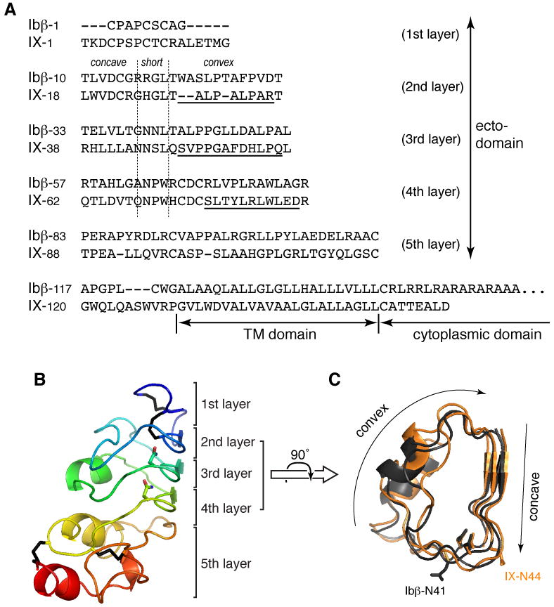
Figure 1. Sequence analysis and structural models of GPIbβ and GPIX ectodomains.

(A) Alignment of human GPIbβ and GPIX sequences. Ectodomain residues are organized into rows corresponding to the 5 layers of the LRR domain. Vertical dashed lines mark the junctions between concave, short and convex sides of the domain. (B) Structural model of the GPIbβ ectodomain, shown in the ribbon diagram with rainbow coloring. The 5-layer organization of the domain is marked on the right. Four disulfide bonds are colored black; side chains of stacked Asn residues in LRR motifs are also shown. (C) A top view of superimposed 2-4 layers of GPIbβ (dark gray) and GPIX (light orange) ectodomains. Concave and convex surfaces are marked. Side chains of Asn ladder point to the inside, while those of glycosylated Asn residues (Ibβ-N41 and IX-N44) point outside.
