
Figure 1.
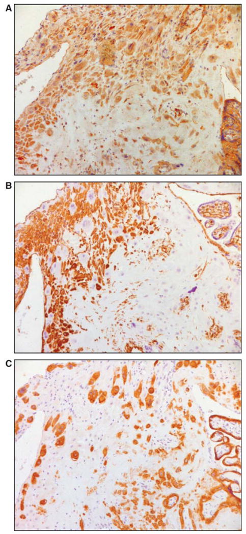
Immunohistochemical analysis of decidual CSF2-expressing cells. Serial sections of a placental bed specimens were stained for CSF2 (A), vimentin (B), and cytokeratin (C). Immunohistochemistry was performed by the labelled streptavidin–biotin (LSAB) method; ×200 original magnification. CSF2 = colony stimulating factor 2.