
Fig. 4.
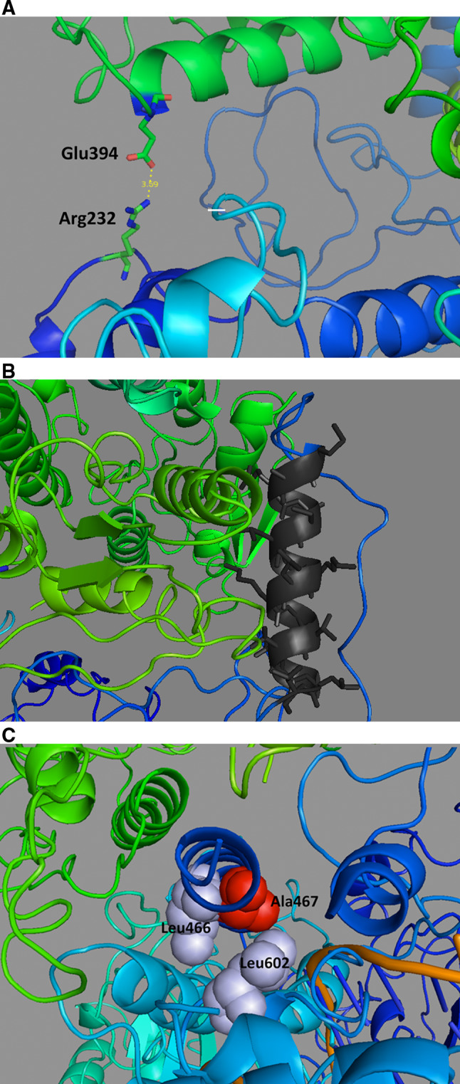
The 3-dimensional structure of the human DNA polymerase γ holoenzyme depicting several residues involved in disease and important for interaction between the catalytic pol γ-α subunit (blue) and the accessory pol γ-β subunit (green). These illustrations were derived using PDB 3IKM [74] in the program PyMOL (http://www.pymol.org/). a The salt bridge between Arg232 of the pol gamma catalytic subunit and Glu394 of the distal p55 accessory subunit. b Pol γ- α amino acids 543–558 (shown in black) form a helix of hydrophobic residues that stabilize interactions with the proximal pol γ- β subunit. c Ala467 is located near Leu466 and Leu602, which is postulated to comprise an important hydrophobic environment for subunit interaction